Radiology – MRI, CT, X-ray, Ultrasound, Mammography
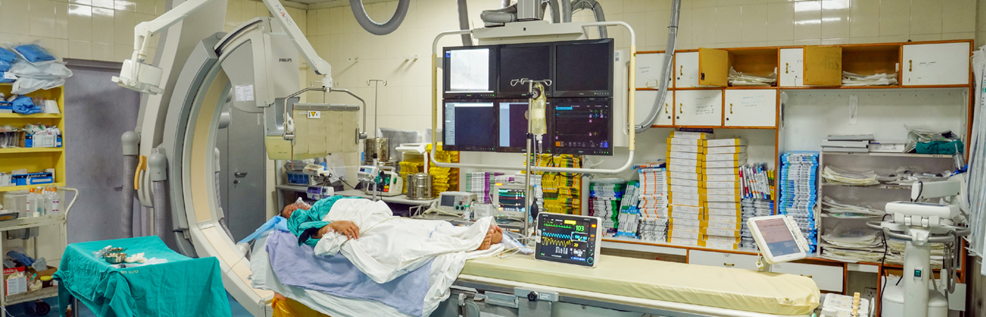
Radiology is a branch of medicine that makes use of imaging technology to diagnose and treat diseases. Its two divisions in the main are Diagnostic and Interventional. Experts who specialize in radiology are referred to as radiologists.
A variety of imaging techniques such as radiography, ultrasound, computed tomography (CT), and magnetic resonance imaging (MRI) are used to diagnose and/or treat diseases. Interventional radiology is the performance of (minimally invasive) medical procedures with the guidance of imaging technologies.
Our Team

Dr. Shailendra Raghuvanshi
Professor & Head

Dr. D. N. Awasthy
Professor Emeritus

Dr. Garima Shailendra Sharma
Assistant Professor

Dr. Gaurav Benjwal
Assistant Professor

Dr. Mamta Goyal
Professor

Dr. Manju Saini
Professor

Dr. Mohan Singh Kunwar
Professor Emeritus

Dr. Satish Chandra Uniyal
Professor

Dr. Vinayak Madhukar Jedhe
Assistant Professor

Dr. Prachi Kala
Professor
-
MRI
Magnetic Resonance Imaging (MRI) is a medical imaging technique that uses a powerful magnetic field, radio waves, and a computer to generate detailed images of the body's internal structures.
The MRI machine itself is a large, cylindrical device that houses a strong magnet. The patient lies on a table that slides into the opening of the machine, and the magnet generates a powerful magnetic field that aligns the protons in the patient's body. Radio waves are then used to stimulate the protons, causing them to emit signals that are detected by the machine's sensors. A computer to generate highly detailed images of the patient’s internal structures then processes these signals.
-
CT
Computed Tomography (CT) is a medical imaging technique that uses X-rays and advanced computer software to create detailed, cross-sectional images of the body's internal structures. CT scanners are are used to diagnose a wide range of medical conditions.
The CT machine is a large, circular device that houses an X-ray tube and a detector. The patient lies on a table that slides into the opening of the machine, and the X-ray tube rotates around the patient, emitting a series of X-ray beams. The detector records the amount of radiation that passes through the patient's body and sends this information to a computer, which uses it to generate detailed images of the patient's internal structures.
-
X-ray
X-ray imaging is a medical diagnostic technique that uses ionizing radiation to create images of the body's internal structures. X-ray machines are commonly found in hospitals and clinics and are used to diagnose a wide range of medical conditions.
The X-ray machine itself is a large, rectangular device that houses an X-ray tube and a detector. The patient lies on a table or stands in front of the machine, and the X-ray tube emits a focused beam of radiation through the body. The detector records the amount of radiation that passes through the patient's body and sends this information to a computer, which uses it to create images of the internal structures.
-
Ultrasound
Ultrasound imaging, also known as sonography, is a medical diagnostic technique that uses high-frequency sound waves to create images of the body's internal structures. Ultrasound machines are commonly found in hospitals and clinics and are used to diagnose a wide range of medical conditions.
The ultrasound machine itself is a small, handheld device that houses a transducer, which emits high-frequency sound waves. The patient lies on a table, and a gel is applied to the skin over the area being examined. The transducer is then moved over the skin, emitting sound waves that bounce off the internal structures and create images that are displayed on a computer screen.
-
Mammography
Mammography is a specialized type of medical imaging that uses low-dose X-rays to create detailed images of the breast tissue. Mammography machines are commonly found in hospitals and clinics and are used to screen for and diagnose breast cancer.
The mammography machine itself is a large, specialized X-ray machine that houses an X-ray tube and a detector. The patient's breast is compressed between two plates, and the X-ray tube emits a focused beam of radiation through the breast tissue. The detector records the amount of radiation that passes through the breast tissue and sends this information to a computer, which uses it to create images of the internal structures.
Mammography can be used either for screening or for diagnostic purposes in evaluating a breast lump :
- Screening mammography - Screening mammography is used to detect signs of breast cancer in women who have no signs or symptoms or new breast abnormalities.
- Diagnostic mammography - Diagnostic mammography is used to investigate suspicious breast changes, such as a new breast lump, breast pain, an unusual skin appearance, nipple thickening or nipple discharge. It is also used to evaluate abnormal findings or if a change is seen on a screening mammogram.
Some general guidelines for when to begin screening Mammography :
- Women with an average risk of breast cancer - Women may begin mammograms at the age of 40 and have them every one to two years. The American Cancer Society advises women with an average risk of cancer to begin screening mammograms yearly at the age of 45 until the age of 54, and then continue every two years.
- Women with a high risk of breast cancer - Women with a high risk of breast cancer may benefit by screening mammograms before the age of 40 after consultation with the doctor about evaluation of an individual’s risk of breast cancer. The risk factors, such as a family history of breast cancer or a history of precancerous breast lesions, may suggest recommendation of Magnetic Resonance Imaging (MRI) in combination with mammograms.